| |
ELG 7173 - Assignment #1
- X-ray imaging: Beam Hardening
Consider a parallel beam X ray source which outputs
the following energy distribution
| Number of Photons
| Photon Energy
|
106
| 10 keV
| |
105
| 20 keV
| |
104
| 30 keV
| |
103
| 40 keV
| |
102
| 50 keV
| |
The X-rays travel through a mixture of soft tissue
and bone. At these frequencies, these tissues have
the following attenuation coefficients:
| Photon Energy
| Attenuation Coefficient
of Bone
| Attenuation Coefficient
of Soft Tissue
|
| 10 keV
| 2.7 cm-1
| 0.6 cm-1
|
| 20 keV
| 0.43 cm-1
| 0.25 cm-1
|
| 30 keV
| 0.28 cm-1
| 0.20 cm-1
|
| 40 keV
| 0.24 cm-1
| 0.19 cm-1
|
| 50 keV
| 0.20 cm-1
| 0.18 cm-1
|
X-rays are emitted from a parallel ray source toward the target
in figure 1. Measurements A and B represent the
energy of photons captured after travelling through the target.
The contrast is ( A − B ) / B.
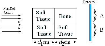
Figure 1: X-ray source, target and detector
-
(5 Marks)
Two different objects are measured. Object #1 has
d1=9 cm, and
d2=1 cm. Object #2 has
d1=4 cm, and
d2=1 cm.
What is the contrast for each object?
-
(5 Marks)
Briefly explain the effect of "beam hardening"
in your result?
- X-ray imaging #2
-
(5 Marks)
Bone is more dense than soft tissue and has a significantly
larger composition in heavier chemical elements
(e.g.
Heymsfield et al, (1991) "Chemical and elemental analysis of humans in
vivo using improved body composition models", AJP - Endocrinology and
Metabolism, 261(2):E190-E198
)
Describe how each of these effects results in
bone having a higher X-ray attenuation than soft tissue
-
(5 Marks)
One way to increase contrast in X-ray images is to
inject contrast agents, such as iodine, into the
blood. For example
Angiography
images are done this way.
Does iodine have higher or lower attenuation than soft tissue?
Why?
- Computed Tomography
The following function makeproj calculates
CT projections from matrix image data.
    function proj= makeproj( a, x, y)
        dointerpolate=1;

        if ~all(diff([size(x),size(y)])==0);
           error('x and y must be square');
        end

        rmax= max([abs(x(:));abs(y(:))]);
        spc = max(abs([mean(mean(diff(x'))), mean(mean(diff(y ))) ]));
        plen= size(x,1);

        %create x indices of projection ray
        xx=x*sin(a) + y*cos(a);
        xidx= (xx + rmax) / spc + 1;
        xi_l=  floor(xidx);     xi_h =  xi_l+1;
        xi_il= (xi_h-xidx);     xi_ih= (xidx-xi_l);

        %create y indices of projection ray
        yy=x*cos(a) - y*sin(a);
        yidx= (yy + rmax) / spc + 1;
        yi_l=  floor(yidx);     yi_h =  yi_l+1;
        yi_il= (yi_h-yidx);     yi_ih= (yidx-yi_l);

        % keep elements within bounds
        kp = (xx.^2 + yy.^2) < (rmax - spc);
        pnum = ones(plen,1) * (1:plen); pnum = pnum(kp);

        % create sparse approximation matrix
    if dointerpolate
        posn =  [ yi_l(kp), yi_h(kp), yi_l(kp), yi_h(kp)] + ...
          plen*([ xi_l(kp), xi_l(kp), xi_h(kp), xi_h(kp)]-1);
        aprx =  [xi_il(kp).*yi_il(kp), xi_il(kp).*yi_ih(kp), ...
                 xi_ih(kp).*yi_il(kp), xi_ih(kp).*yi_ih(kp)];
        proj = sparse( [pnum,pnum,pnum,pnum], posn, aprx,plen,plen^2);
    else
        xi_r= round(xidx); yi_r= round(yidx);
        posn =  yi_r(kp) + plen*(xi_r(kp)-1);
        proj = sparse( pnum, posn, 1 ,plen,plen^2);
    end
|
A sample usage of this code to implement (non-filtered)
simple backprojection is as follows:
    spc=.025; rlim=1;
    [x,y]= meshgrid(-rlim:spc:rlim,-rlim:spc:rlim);
    plen= size(x,1);
    img = (x.^2 + y.^2) > rlim;
    img( x >.45 & x<.65 & y>-.05 & y<.45) =1;
    img( x >-.55 & x<-.25 & y>.45 & y<.65) =1;

    i=1; proj= zeros(plen,6);
    for ang= [0:30:150];
      prm= makeproj(ang*(pi/180),x,y);
      proj(:,i) = prm* img(:);
      i=i+1;
    end

    imbp= zeros(size(img));
    i=1;
    for ang= [0:30:150];
      prm= makeproj(ang*(pi/180),x,y);
      imbp= imbp + reshape( prm' * proj(:,i) , plen, plen);
      i=i+1;
    end
    imbp = imbp + max(imbp(:))*(  x.^2 + y.^2 > rlim );

    imagesc(imbp);
|
Using this sample code, we investigate the noise properties of
simple backprojection and filtered backprojection.
- (5 Marks)
Increase the number of projection angles.
Show at least two images of low and high quality
images
Comment on the improvement in image quality with number of
projections. Roughly, at what number of projections,
does the quality improvement become less apparent?
- (15 Marks)
Implement the "filtered backprojection"
or "convolution backprojection" algorithm (your choice),
by modifying the given code to prefilter the projection data.
If you wish, you may also implement any other CT reconstruction
although this will be more difficult.
Comment on the improvement in image quality with
your implemented technique. Are there any
noise problems with this technique?
- Computed Tomography #2
Consider the geometry defined in the following figure for a
model of a computed tomography system. Seven hexagonal
regions, R1 to R7 are defined.
Each region is 2 cm wide between parallel faces.
Regions R1, R2, R3
R5, R6, and R7
are soft tissue, and
region R4 is bone.
The attenuation coefficient for
bone is 1.2 cm−1, and for
soft tissue is 0.5 cm−1.
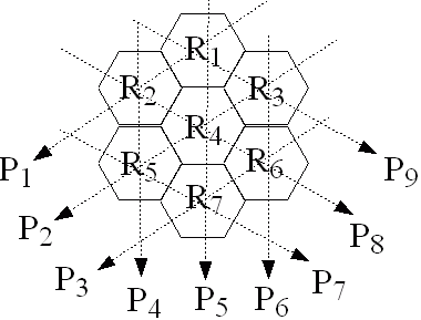
Figure: Geometry for CT system
- (5 marks)
Each projection has 106 input X-ray
photons. Define each projection to
be P=½ln( Nin/Nout)
Calculate P1 ... P9
- (5 marks)
Write the matrix equation relating the
vector of region attenuations
( R1 ... R7)
to the vector of projections
( P1 ... P9)
- (5 marks)
Write a function to calculate the region
attenuations using ART. How accurate
is your estimate after three iterations?
Last Updated:
$Date: 2007-01-22 14:44:23 -0500 (Mon, 22 Jan 2007) $
|