Contributed EIT Data:

Authors: I. Frerichs, G. Hahn, T. Schröder, G. Hellige
Date: 1998
Brief Description: Five pigs before (e.g. 1-control.raw) and after induction of unilateral lung injury (e.g. 1-injury.raw). Oleic acid was administered through the distal opening of a pulmonary artery catheter placed in a branch of the left pulmonary artery.

Data were published in:
I. Frerichs, G. Hahn, T. Schröder, G. Hellige Electrical impedance tomography in monitoring experimental lung injury Intensive Care Med. 24:829-836, 1998.


License: Creative Commons Artistic License (with Attribution)
Attribution Requirement: Use or presentation of these data must acknowledge Inéz Frerichs, and cite this publication:
Format: EIT data were acquired with the Sheffield APT device Mk 1. Measurement duration 240 s, frame rate 1 Hz.
Methods: Five pigs before (e.g. 1-control.raw) and after induction of unilateral lung injury (e.g. 1-injury.raw). Oleic acid was administered through the distal opening of a pulmonary artery catheter placed in a branch of the left pulmonary artery.
Ventilator settings:
Volume-controlled ventilation
PEEP: 5 cm H2O
FIO2: 0.5
Fresp: 12 /min
The ventilator settings were not changed!

Data: Data (zip format)
figure 3
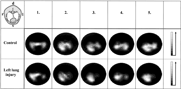
Figure 3 (from Frerichs et al, 1998): Thoracic functional EIT images before (Control) and after the development of artificially induced left lung injury (Lung injury) reflecting the distribution of ventilation in the five animals studied. (The lungs are typically located in the central and ventral parts of the thorax due to the well developed backbone musculature in pigs.) The schematic cross-section of the pig thorax in the left upper corner shows the spatial orientation of the images. Each image is scaled to the individual maximum impedance variation. The scale shows that higher local impedance variation (i.e. higher fluctuation of regional lung volume) is represented in light tones

Last Modified: $Date: 2017-02-28 13:12:08 -0500 (Tue, 28 Feb 2017) $ by $Author: aadler $