| Authors: |
G. Hahn, A. Just, T. Dudykevych, I. Frerichs, J. Hinz, M. Quintel and G. Hellige
| ||||||||||
|---|---|---|---|---|---|---|---|---|---|---|---|
| Date: | 2006
| ||||||||||
| Brief Description: |
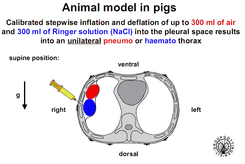
Studies on five anaesthetized
(Fentanyl/Midazolam) mechanically ventilated supine pigs
(mean body weight 30.1 ± 2.3 kg). Tidal volume (15 ml/kg)
and ventilation frequency were kept constant in
each animal for all tomograms during the experiments. To
cause controlled air or fluid accumulation, up to 300
ml of air or 300 ml of Ringer solution was injected
unilaterally at the right side into or from the pleural
space in steps of 100 ml. The first injection was always air
since Ringer solution could not be completely removed in
most cases. A small incision was made in the chest wall
and a plastic canula was fixed by a suture and
additionally sealed by cyanoacrylate glue. Sixteen ECG
electrodes (Blue Sensor, VL00S, Medicotest, Olstykke, DK)
were placed around the thorax 3 cm below the axilla. The
reference electrode was placed approximately 10 cm below
the electrode plane. Series of EIT measurements of 30 s
duration were performed at a rate of 13 frames per second
using a Goe-MF II tomography system (Hahn et al 2002). An
adjacent current injection and measurement pattern was
used. The frequency of the injected current was 50 kHz at
an amplitude of 5 mArms. EIT measurements were performed
either after complete injection of 300ml air or Ringer
solution. In two pigs, themeasurements were performed
during the whole period of stepwise injection. In each
animal, air was injected first and completely removed
before Ringer solution was injected. The whole protocol
was approved by the State Animal Care Committee.
Data were published in G Hahn, A Just, T Dudykevych, I Frerichs, J Hinz, M Quintel and G Hellige (2006) Imaging pathologic pulmonary air and fluid accumulation by functional and absolute EIT Physiol. Meas. 27:S187−S198 | ||||||||||
| License: | Creative Commons Artistic License (with Attribution)
| ||||||||||
| Attribution Requirement: |
Use or presentation of these data must acknowledge
Günter Hahn, and reference this publication:
| ||||||||||
| Format: | Data are in *get files encoded in a zip file
| ||||||||||
| Methods: | Pig torso. Single plane of
16 Electrodes numbered clockwise, with electrode #1 at the top.
| ||||||||||
| Data: | Data (zip format)
|
Last Modified: $Date: 2017-02-28 13:12:08 -0500 (Tue, 28 Feb 2017) $ by $Author: aadler $