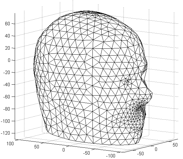
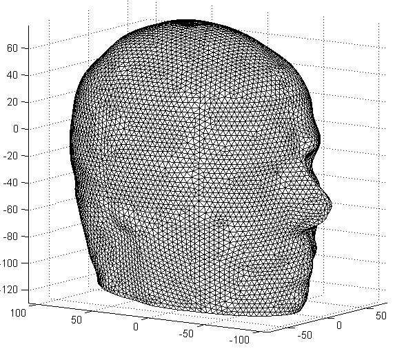
Figure: Left: Outer boundary of SAH028 (27684 element) mesh Right: Outer boundary of SAH262 (262368 elements) mesh Last Modified: $Date: 2017-02-28 13:12:08 -0500 (Tue, 28 Feb 2017) $ by $Author: aadler $
| Authors: |
Andrew Tizzard,
Middlesex University, Bramley Road, London N14 4YZ.
| ||||||||||||||||||||||||||||||||||||||||||||||||||||||||||||||||||
|---|---|---|---|---|---|---|---|---|---|---|---|---|---|---|---|---|---|---|---|---|---|---|---|---|---|---|---|---|---|---|---|---|---|---|---|---|---|---|---|---|---|---|---|---|---|---|---|---|---|---|---|---|---|---|---|---|---|---|---|---|---|---|---|---|---|---|---|
| Date: | October 2007. (Created from 2002 to 2006)
| ||||||||||||||||||||||||||||||||||||||||||||||||||||||||||||||||||
| Brief Description: |
A range Finite Element meshes of different densities for the human
head including two neonate meshes.
The underlying geometry is described in Tizzard et al. (2005b) and all meshes except SA262 are used in Tizzard & Bayford (2007a) | ||||||||||||||||||||||||||||||||||||||||||||||||||||||||||||||||||
| License: | Creative Commons Artistic License (with Attribution)
| ||||||||||||||||||||||||||||||||||||||||||||||||||||||||||||||||||
| Attribution Requirement: |
Tizzard, A. and Bayford, R.H. (2007).
Improving the Finite Element Forward Model of the Human Head by
Warping using Elastic Deformation.
Physiol.Meas. 28:S163-S182.
| ||||||||||||||||||||||||||||||||||||||||||||||||||||||||||||||||||
| Format: |
Data are in Matlab v6 format,
compressed with 7-Zip
Each model is shelled comprising regions defining Scalp, Skull, CSF and brain as defined by the materials structure. The important arrays in this structure are names and ref. The vector ref relates elements (tri) to each region by index into the names array. Nodal positions (mm) are defined by the vtx array. The underlying geometry is described in Tizzard et al. (2005b) and all meshes except SA262 are used in Tizzard and Bayford (2007a) | ||||||||||||||||||||||||||||||||||||||||||||||||||||||||||||||||||
| Methods: |
Geometry generated using Alias Studio Tools (now Autodesk Alias)
and the meshing carried out using EDS I-DEAS (now UGS NX).
The tables below give a summary of the adult and neonate head meshes. They are all shelled comprising regions defining Scalp, Skull, CSF and brain. The underlying geometry is described in Tizzard et al. (2005b) and all meshes except SA262 are used in Tizzard and Bayford (2007a) Some of these meshes and others based on the same underlying geometry have been used by the UCL and Middlesex groups (Bagshaw et al. 2003; Bayford et al. 2005; Bayford et al. 2006b; Bayford et al. 2006a; Horesh et al. 2004; Tizzard et al. 2005a; Tizzard and Bayford 2007b; Yerworth et al. 2004).
Summary of shelled adult head meshes.
Summary of shelled neonate head meshes
References
| ||||||||||||||||||||||||||||||||||||||||||||||||||||||||||||||||||
| Data: |
Adult head meshes:
−SAH028 (27684 elements), −SAH031 (31111 elements), −SAH035 (35073 elements), −SAH049 (49172 elements), −SAH077 (77196 elements), −SAH132 (131672 elements), −SAH262 (262368 elements) Neonate head meshes: −SNH046 (45702 elements) −SNH143 (142654 elements) |
 for str = {'SAH028', 'SAH262'}
    load(str{1});
    ee= find_boundary(tri);
    trimesh(ee,vtx(:,1),vtx(:,2),vtx(:,3))
    hh=trimesh(ee,vtx(:,1),vtx(:,2),vtx(:,3));
    set(hh,'EdgeColor', [0,0,0]);
    axis tight
    axis equal
    view(-55,10);
    print('-dpng','-r100',[str{1},'.png']);
end